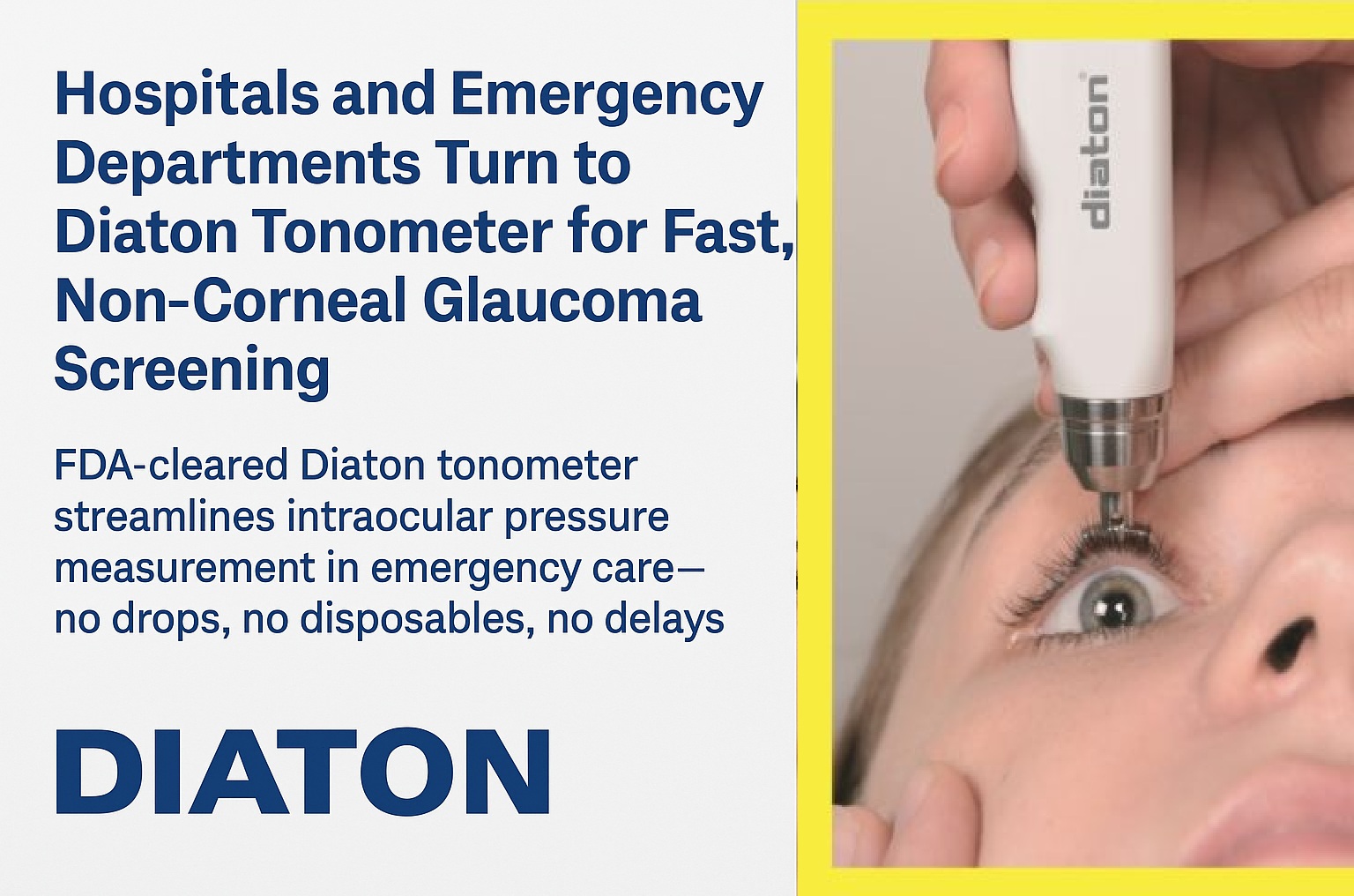
Intraocular pressure can provide important clinical information in emergency care. But when IOP is measured through the cornea, an important question often goes unasked: how much is the cornea itself affecting the number?
In ophthalmology, clinicians routinely think about central corneal thickness (CCT), corneal biomechanics, previous refractive surgery, edema, scarring, ectasia, and other factors when interpreting intraocular pressure.
In the emergency department, the situation can be very different.
A patient may arrive with eye pain, redness, headache, visual changes, trauma, or another ocular complaint. IOP may be clinically relevant, yet the patient’s complete ophthalmic history may be unavailable. Pachymetry may not be readily available. And the clinician obtaining the pressure measurement may have no reason to know whether the patient’s cornea is unusually thin, thick, surgically altered, irregular, edematous, or otherwise abnormal.
That raises an important question:
How much do you know about the cornea behind the IOP number?
Goldmann applanation tonometry remains the clinical gold standard for IOP measurement in ophthalmology. It estimates IOP by measuring the force required to flatten a defined area of the cornea. (EyeWiki)
That measurement is therefore not completely independent of the properties of the cornea being measured.
Central corneal thickness is one important variable. In general, thicker corneas can produce artificially higher Goldmann readings and thinner corneas can produce artificially lower readings. But CCT is only part of the story. Corneal elasticity, rigidity, curvature, astigmatism, edema, scarring, ectasia, tear-film characteristics and previous surgery can also affect corneal-dependent measurements. (EyeWiki)
Importantly, this isn’t simply a problem that can always be solved by measuring CCT and applying a mathematical correction.
AAO EyeWiki notes that the effect of CCT on IOP is not completely predictable and that there is no consensus regarding a universal degree of correction for thin or thick corneas. CCT is only one biomechanical characteristic of the cornea. (EyeWiki)
For an ophthalmologist, these considerations are familiar.
For an emergency physician, nurse, technician, urgent-care provider, or other clinician outside dedicated eye care, interpreting corneal biomechanics is understandably not the primary job.
Consider a seemingly ordinary IOP reading
Suppose a patient presents to the ED and a corneal-dependent tonometer produces an IOP of 17 mmHg.
The number by itself may appear unremarkable.
But what else do we know?
Has the patient previously undergone LASIK or PRK?
Is the cornea unusually thin or thick?
Does the patient have early keratoconus or another ectasia?
Is there corneal edema?
Scarring?
Irregular astigmatism?
Previous corneal surgery?
The person measuring IOP may not know.
And the patient may not volunteer that history, particularly during an emergency encounter.
This does not make the IOP measurement useless. It means that the measurement method and the condition of the cornea are part of the clinical context in which that number should be interpreted.
LASIK provides a good example
Millions of people have undergone corneal refractive procedures.
LASIK changes central corneal thickness and corneal biomechanics. AAO EyeWiki notes that these changes can result in artifactually low IOP measurements with Goldmann applanation tonometry. Similar measurement issues have been documented following PRK and other refractive procedures. (EyeWiki)
Years later, a patient presenting to an emergency department may simply say:
“I had laser eye surgery a long time ago.”
Or they may not mention it at all.
Yet that altered cornea can still matter to interpretation of a corneal-dependent pressure measurement.
This illustrates a broader issue:
The emergency clinician may be measuring not only IOP, but IOP through a biological structure whose characteristics are unknown.
Pachymetry helps — but doesn’t tell the entire story
Central corneal thickness is valuable clinical information, and pachymetry has an important role in ophthalmic evaluation.
But thickness alone does not completely describe corneal biomechanics.
Two corneas with similar thickness can behave differently mechanically. Conversely, pathological thickening from edema does not necessarily behave like a naturally thick, healthy cornea. AAO’s review of IOP reliability describes how physiologically and pathologically thick corneas can affect measurements differently. (EyeWiki)
That is why simply asking:
“What is the patient’s CCT?”
doesn’t necessarily answer:
“How much is this patient’s cornea affecting this IOP measurement?”
For an emergency department attempting to establish practical bedside IOP capability, that distinction matters.
What if you take the cornea out of the equation?
There is another approach to tonometry.
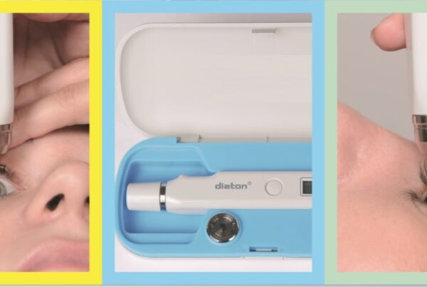
Instead of measuring IOP through the cornea, transpalpebral scleral tonometry measures through the upper eyelid in the scleral region.
Diaton uses this different measurement approach.
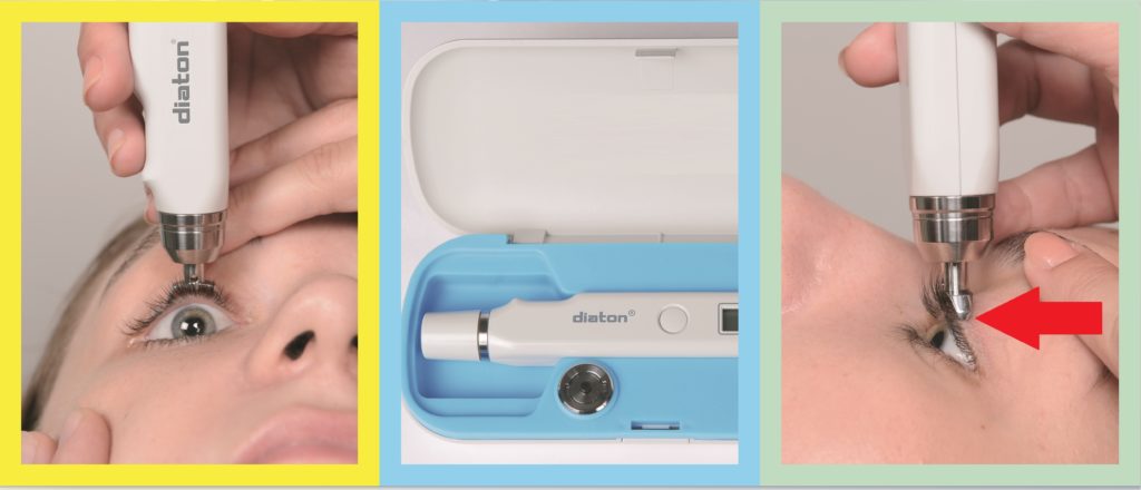
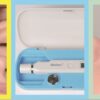
The FDA-cleared Diaton Tonometer is intended to measure intraocular pressure as an aid in the diagnosis of glaucoma and for monitoring IOP. Its FDA documentation describes IOP measurement through the eyelid in the scleral area, without requiring anesthetic for the measurement. (FDA Access Data)
Because the measurement does not use the cornea as its measurement surface, central corneal thickness is not an input required to obtain the Diaton measurement.
That distinction may be particularly relevant outside traditional ophthalmology environments.
It doesn’t mean that the condition of the cornea is clinically irrelevant. Corneal pathology may itself require diagnosis and treatment, and no tonometer replaces a complete eye examination or ophthalmologic consultation when indicated.
Rather, it means that the IOP measurement itself is obtained using another anatomical route.
Why might that matter in emergency medicine?
For a hospital or emergency department, the potential value extends beyond corneal biomechanics.
A practical IOP solution also has to fit the environment in which clinicians actually work.
Diaton’s FDA documentation describes a handheld device capable of measurement with patients sitting or supine, with a single measurement taking no more than three seconds under its specified operating conditions. It does not require topical anesthesia for the measurement. (FDA Access Data)
That creates a different workflow proposition:
Portable IOP measurement.
No corneal contact.
No anesthetic drops required for the Diaton measurement.
No CCT measurement required as an input to obtain the Diaton reading.
Sitting or supine measurement.
For an ophthalmology practice, these may be conveniences or complementary capabilities.
For a setting that does not have a complete ophthalmic lane, they may represent something more fundamental:
greater practical access to IOP measurement.
Diaton is not a substitute for ophthalmology
This distinction is important.
A tonometer does not diagnose glaucoma by itself. Nor should an IOP number alone determine the management of an ocular emergency.
IOP should be interpreted alongside symptoms, history, examination findings and the broader clinical picture. Patients requiring specialist evaluation should still receive appropriate ophthalmologic consultation or referral.
And different tonometry technologies have their own sources of variability and technique requirements.
The purpose of a different measurement approach is not to declare established tonometry obsolete.
It is to provide clinicians with another way to obtain IOP information when measuring through the cornea may be undesirable, difficult, or potentially affected by corneal characteristics.
A different question for emergency departments
When hospitals evaluate tonometry equipment, the conversation often starts with:
“Which tonometer should we buy?”
Perhaps the better questions are:
Who needs to measure IOP?
Where does the measurement need to happen?
What additional equipment and consumables does the workflow require?
How much training is required?
Does the measurement depend on corneal characteristics that may be unknown?
Can the device be used where the patient already is?
Those questions move the discussion beyond device specifications and toward actual clinical workflow.
And they lead to one final question:
How much is the cornea affecting your IOP reading?
When the answer is uncertain, it may be useful to have another perspective.
Diaton — IOP through the eyelid and sclera.
Think Beyond the Cornea.™
Evaluating IOP measurement options for your Emergency Department?
Request information about the Diaton Hospital & Emergency Medicine Evaluation Program.
Contact Team Diaton: www.TonometerDiaton.com / 1-877-342-8667

FAQ
Does corneal thickness affect IOP measurements?
Corneal thickness can influence IOP measurements obtained with corneal-dependent tonometry. In general, unusually thick or thin corneas can affect measured IOP, but corneal thickness is only one factor. Corneal biomechanics, curvature, disease, previous surgery and other characteristics can also influence measurement and interpretation.
Can LASIK or PRK affect an IOP reading?
Yes. Refractive procedures such as LASIK and PRK alter corneal thickness and biomechanics and can affect IOP measurements obtained through the cornea. A patient’s history of refractive surgery should therefore be considered when interpreting corneal-dependent tonometry.
Does pachymetry correct an IOP reading?
Pachymetry provides central corneal thickness, which can be important when interpreting IOP. However, CCT alone does not characterize all aspects of corneal biomechanics, and there is no universally accepted correction formula that converts every corneal-dependent measurement into a “true” IOP.
What other corneal conditions can complicate IOP measurement?
Keratoconus and other ectasias, corneal edema, scarring, irregular corneal surfaces, keratoprosthesis, previous refractive or corneal surgery and other structural changes can complicate some methods of IOP measurement or their interpretation.
What is cornea-independent tonometry?
Cornea-independent tonometry obtains IOP information without using the cornea as the measurement surface. Diaton uses a transpalpebral scleral approach, measuring through the upper eyelid in the scleral region rather than through the cornea.
Does Diaton require pachymetry?
Central corneal thickness is not an input required to obtain a Diaton IOP measurement because Diaton does not measure through the cornea. Pachymetry may still be clinically indicated for other aspects of a patient’s ophthalmic evaluation.
Does Diaton touch the cornea?
No. Diaton measures through the upper eyelid over the scleral region and does not contact the cornea.
Are anesthetic eye drops required with Diaton?
Topical anesthetic eye drops are not required to obtain a Diaton IOP measurement.
Can Diaton be used with a patient lying down?
Diaton can be used with appropriately positioned patients in sitting or supine positions, which may be useful in hospital and bedside environments.
Is Diaton intended to replace Goldmann applanation tonometry?
Diaton provides a different approach to IOP measurement rather than simply duplicating corneal applanation. The appropriate tonometry method and interpretation of an IOP result depend on the clinical situation. Diaton can provide an additional IOP perspective, particularly when measuring through the cornea presents limitations or challenges.
Can an IOP measurement diagnose glaucoma?
No. IOP is an important clinical measurement and elevated IOP is a major risk factor for glaucoma, but glaucoma diagnosis requires evaluation of the broader clinical picture, which may include the optic nerve, visual fields, imaging, history and other findings.
Why could portable tonometry be useful in an emergency department?
Emergency departments frequently evaluate patients away from dedicated ophthalmic examination rooms. A portable tonometer can make IOP measurement available closer to the patient’s bedside and may reduce dependence on fixed ophthalmic equipment for obtaining the measurement.
For Emergency Physicians, ED Directors, Hospital Administrators & Clinical Educators
Reviewed by Michael Minch